
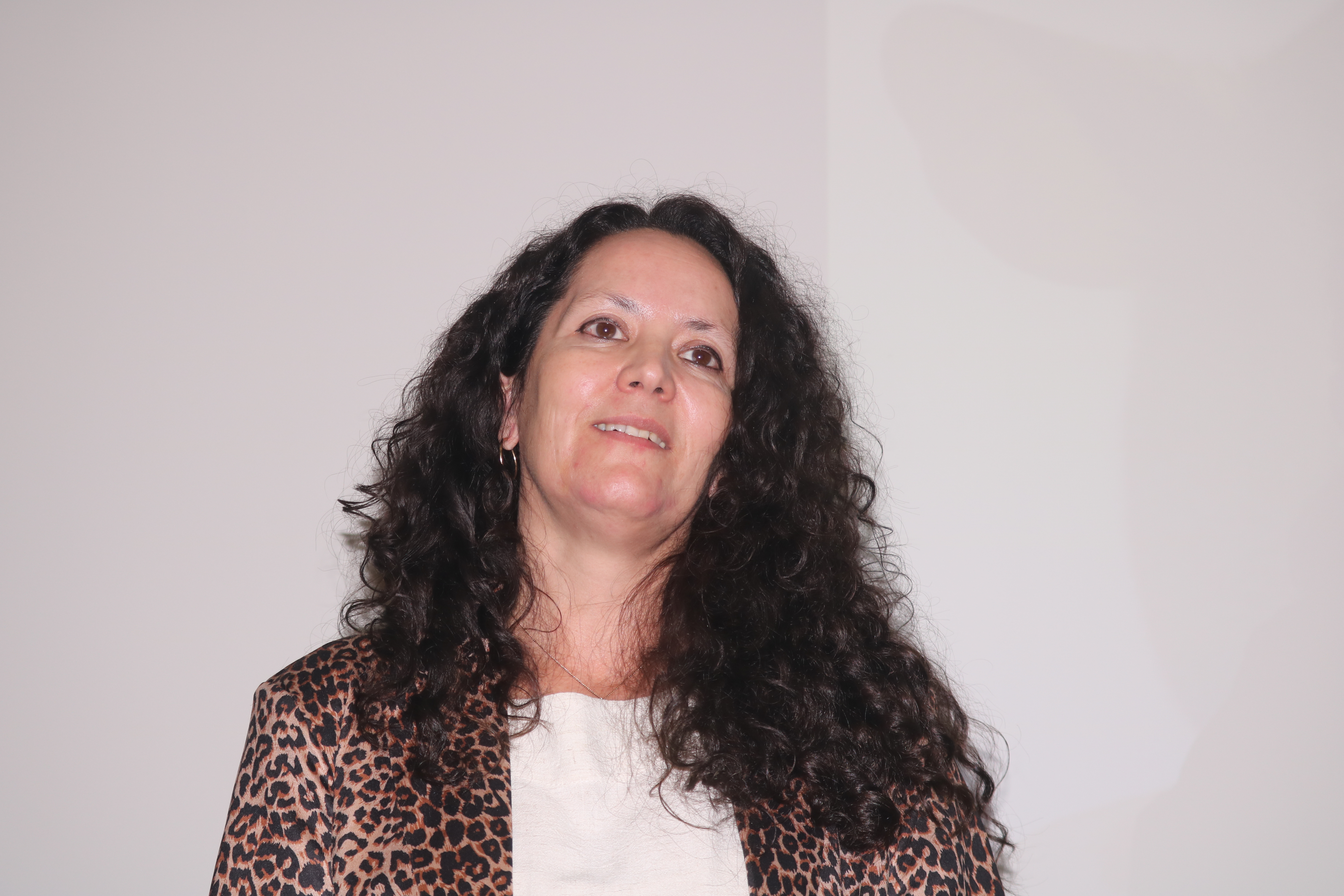
The Animal Biotechnology Unit and the Advanced Bioimaging Unit at the Institut Pasteur de Montevideo were among the highlights of the second day of the IPSP–IPMon meeting, alongside research on Chagas disease, SARS-CoV-2, Zika virus, and organoids applied to the study of infectious diseases.
Discussions on the morning of the second day of the IPSP & IPMon – A Pasteurean Scientific Meeting, held from September 16 to 18, highlighted an essential element of contemporary infectious disease research: the development of platforms capable of reproducing the complexity of biological systems with increasing fidelity. From facilities for generating genetically modified animal models to advanced bioimaging tools, organoids, and molecular analyses, the presentations showed how new research infrastructures are expanding the possibilities for understanding interactions between pathogens and hosts.
Ana Paula Arevalo presented the Laboratory Animal Biotechnology Unit (UBAL) at the Institut Pasteur de Montevideo. The unit brings together technologies for producing genetically modified animal models, developing preclinical models, conducting pharmaceutical testing, and providing specialized training. It currently maintains more than 60 genetically modified lines and produces over 20,000 mice per year.
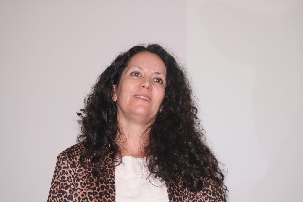
Ana Paula Arevalo (IPMon)
“Among the platform’s capabilities are CRISPR/Cas9 gene editing, embryo manipulation and transfer, in vitro fertilization, cryopreservation, specialized surgical procedures, and in vivo imaging techniques,” said Arevalo.
One of the examples presented was the ongoing development of a personalized experimental model — an “avatar” — based on a genetic alteration identified in a Uruguayan child with a rare disease.
In addition to models for genetic diseases, UBAL also maintains systems for infectious disease research and participated in projects carried out during the Covid-19 pandemic. One of these involved a collaboration with Fiocruz and the University of São Paulo to investigate the localization and biodistribution of fluorescent nanoparticles in an experimental approach related to Covid-19.
Andrés Kamaid Toth presented the Advanced Bioimaging Unit, a facility shared by the Institut Pasteur de Montevideo and the Universidad de la República. The unit combines advanced microscopy, the development of custom-built instruments, computational image analysis, and training programs for researchers across Latin America. In recent years, the platform has served more than 400 users and participated in dozens of scientific collaborations. “The mission of the Advanced Bioimaging Unit is to provide unrestricted access to state-of-the-art bioimaging technologies,” he said.
Toth showed how tools such as confocal microscopy, FLIM (Fluorescence Lifetime Imaging Microscopy), hyperspectral imaging, super-resolution microscopy, and 4D systems make it possible to observe biological phenomena that were previously inaccessible. An important part of the presentation focused on so-called phasor plots, a method that transforms complex fluorescence information into quantitative parameters capable of revealing cellular and metabolic changes.
These technologies are being applied to the study of infections such as those caused by Neospora caninum. Experiments showed changes in cell membrane composition and fluidity, as well as mitochondria-related metabolic alterations during infection. Another example involved the use of zebrafish embryos to monitor immune-cell dynamics in real time and test candidate molecules in models of inflammation and drug discovery.
Congenital transmission of Chagas disease was the focus of Lisvane Paes’s presentation. Her group investigates Trypanosoma cruzi isolates associated with vertical transmission in Uruguay and has identified important differences compared with strains traditionally used in laboratory research. The results indicate that some of these lineages have low virulence and trigger less inflammatory placental responses, suggesting a more silent transmission strategy between mother and fetus. Transcriptomic and proteomic analyses also revealed specific characteristics related to adhesion, invasion, and host interaction.
Also focusing on a technological platform, Mariela Bollati-Fogolin presented intestinal organoids developed by the Cell Biology Unit. Produced from adult stem cells from mice, cattle, and sheep, these models preserve different cell types and structural features of the intestine, allowing infections to be studied in systems that more closely reflect biological conditions.
The organoids are being used in studies involving Trypanosoma cruzi, Salmonella enterica, and Toxoplasma gondii. In experiments on Chagas disease, researchers observed differences in susceptibility to infection among different intestinal regions and showed that co-culture with fibroblasts increased organoid differentiation and reduced susceptibility to infection. In another project, the group succeeded in inducing pre-sexual stages of T. gondii in vitro in murine intestinal organoids, reproducing stages of the parasite’s life cycle that are normally restricted to felines.
Contributions from IPSP
The Institut Pasteur de São Paulo presentations focused mainly on the effects of viral infections and immune responses induced by vaccines. Alba Marina Gimenez presented a vaccine strategy based on recombinant BCG and a chimeric protein against SARS-CoV-2. The experiments showed that CD4+ and CD8+ T-cell responses were required for protection in the model studied, while humoral immunity alone was not sufficient. Protection was maintained against the JN.1 variant despite limited antibody neutralizing activity.
Patrícia Beltrão-Braga presented research using neurons, astrocytes, and brain organoids derived from induced pluripotent stem cells to investigate the impact of Zika virus on neurodevelopment. The studies showed impaired neural progenitor migration, alterations in cortical development, and an important role for the cytokine IL-6 in inflammatory processes also observed in models of autism spectrum disorder.
Closing the morning session, Vanessa Barbosa Malaquias presented a model of human brain organoids maintained for more than 300 days before infection with Zika virus, making it possible to investigate the virus’s effects on more mature neural tissue. Although viral load decreased over time, the organoids continued to show progressive structural loss and increased apoptosis.
The finding supports the hypothesis that a reduction in detectable virus does not necessarily imply recovery of nervous tissue and led to the development of a platform for vaccine evaluation that considers not only viral control but also preservation of brain tissue.