
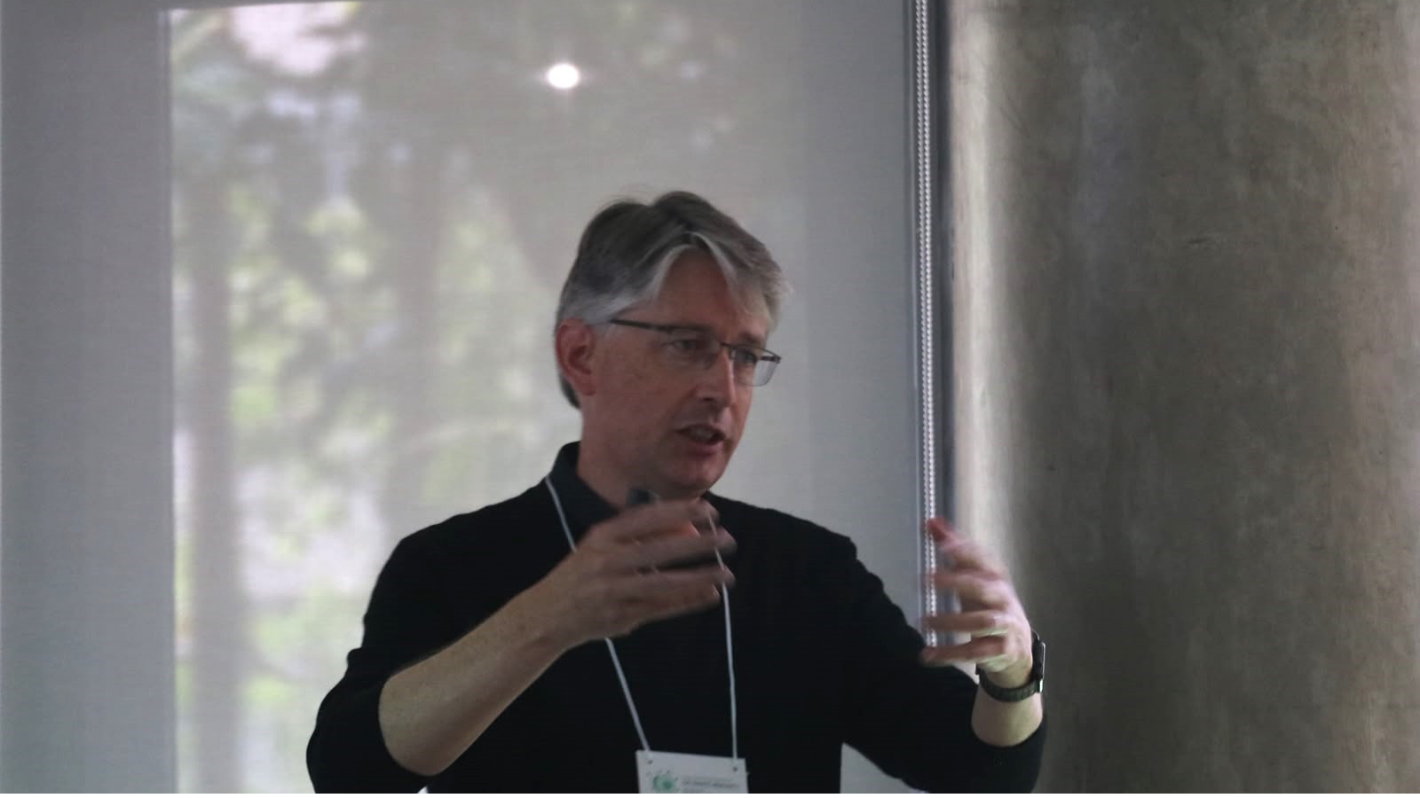
Talks presented on the morning of the final day of the International Symposium on Innate Immunity addressed training the lungs against infections, the use of BCG to enhance antitumor responses, the role of interferons in tuberculosis, and cellular mechanisms involved in controlling pathogens.
How can the body be prepared to respond more effectively to an infection without increasing damage to its own tissues? And what happens when a molecule that is essential to immune defense begins, when produced in excess, to promote disease progression? These two sides of the immune response ran through the presentations held on the morning of September 5, the final day of the International Symposium on Innate Immunity at the University of São Paulo (USP).
The studies showed that previous experiences can reprogram cells and tissues, that BCG can enhance antitumor responses, that a persistent type I interferon response can impair tuberculosis control, and that the internal organization of the phagosome itself influences the ability of macrophages to eliminate pathogens.
The symposium was organized by the University of São Paulo’s Institute of Biomedical Sciences (ICB-USP) and the Institut Pasteur de São Paulo (IPSP), with support from the São Paulo Research Foundation (FAPESP).
Training the immune system—and the lungs as well
Opening the morning program, Maziar Divangahi, from McGill University in Canada, discussed two concepts that guided his presentation: trained immunity and disease tolerance.
The researcher began with a central question: if the adaptive response and its memory are not sufficient to provide effective protection against certain complex infections, could the memory capacity of innate immunity also be harnessed?
Divangahi showed that cells such as monocytes, macrophages, and neutrophils can undergo long-lasting changes following certain exposures, through a process associated with metabolic and epigenetic reprogramming. This phenomenon has become known as trained immunity.
One form of this training occurs in the bone marrow. Stimuli such as BCG vaccination can modify hematopoietic stem and progenitor cells, influencing the characteristics of the immune cells subsequently produced.
The presentation, however, went beyond what is known as central trained immunity. Divangahi also discussed the possibility of training the organ itself, particularly the lungs. Resident and structural cells in lung tissue can retain the effects of previous exposures and respond differently when faced with a new insult.
This idea is connected to the concept of disease tolerance. Surviving an infection more successfully does not always mean eliminating the infectious agent more quickly. In some cases, the determining factor may be the body’s ability to limit the damage caused by its own inflammatory response.
In influenza models, Divangahi presented experiments in which prior treatment with beta-glucan significantly increased survival without producing an equivalent reduction in viral replication. The benefit was associated with less lung damage and the functional reprogramming of immune cells, including neutrophils.
Another focus of the talk was lung tissue recovery. The researcher highlighted the role of type 2 alveolar cells, or AT2 cells, which can proliferate and give rise to AT1 cells, which are essential for rebuilding the alveolar epithelium following injury.
The findings presented indicated that autophagy in AT2 cells is necessary for this repair process. When components of this machinery were eliminated, lung injury and fibrosis increased, while epithelial regeneration was impaired, even without a corresponding increase in viral replication.
The cytokine IL-22 emerged as one of the signals involved in this recovery. The data indicated that its action on AT2 cells promotes mechanisms related to autophagy, cell proliferation, and lung repair.
Divangahi’s message was that preparing the body to face an infection may involve two complementary strategies: improving its ability to control the pathogen and increasing its capacity to preserve and repair the affected tissues.
BCG and the systemic response against cancer
Nina M. G. P. de Queiroz, a researcher in Sergio C. Oliveira’s laboratory at the University of São Paulo’s Institute of Biomedical Sciences, then presented findings on the use of BCG to treat bladder cancer and on the effects of prior immunization on the antitumor response.
To investigate whether the effect could extend beyond the site receiving treatment, the group used an experimental model with two tumors in the same animal and administered BCG directly to only one of them.
The treated tumor was controlled, but a response was also observed in the second tumor, which had not received BCG directly. When the animals had previously been immunized with BCG, greater control was observed in both tumors.
Analysis of the tumor microenvironment revealed changes in different immune cell populations, including macrophages, neutrophils, and T cells. Significant changes were also observed in the tumors’ gene expression profiles following the combination of prior immunization and treatment.
Bone marrow-derived macrophages from previously immunized animals also exhibited metabolic and functional changes, including greater glycolytic capacity and increased cytokine production following renewed stimulation.
The findings therefore point to the possibility that prior exposure to BCG modifies the immune system’s response and subsequently enhances tumor control, including in a tumor that did not receive the treatment directly.
When an intense type I interferon response promotes tuberculosis
The third speaker of the morning, Russell E. Vance, from the University of California, Berkeley, presented an apparently paradoxical perspective: in tuberculosis, a very intense immune response may, under certain conditions, contribute to the progression of the disease itself.
In his talk, “Tuberculosis as an Interferonopathy,” Vance discussed evidence that excessive and persistent levels of type I interferon can increase susceptibility to infection with Mycobacterium tuberculosis.
The researcher noted that only a proportion of infected individuals develop active tuberculosis and that patients with the disease exhibit a strong type I interferon-associated signature. Follow-up studies have also indicated that this signature can emerge before progression to active disease, raising the question of whether it is merely a consequence of the infection or plays a role in its progression.
To investigate this question further, the group used animal models that were genetically more susceptible to tuberculosis. The absence of the SP140 protein was associated with an exacerbated type I interferon response and greater susceptibility to infection. When this interferon signaling was disrupted, a significant part of that susceptibility was reversed.
One of the mechanisms presented involves IL-1, which is important for controlling tuberculosis. Excessive type I interferon can increase the expression of the IL-1 receptor antagonist, thereby reducing this protective signaling.
The group also identified another mechanism. Type I interferon acts on macrophages and other myeloid cells, impairing their ability to respond to interferon gamma, one of the main molecules involved in activating macrophages against the bacterium.
In laboratory experiments, macrophages stimulated with interferon gamma showed strong activation of genes associated with the protective response. When the cells were first exposed to interferon beta, this subsequent response to interferon gamma was almost completely blocked. The effect persisted even after interferon beta was removed.
Vance emphasized that the problem is not the absence of interferon gamma. The molecule continues to be produced. What is lost is the ability of infected cells to respond to it appropriately.
The data led the researcher to propose that, in certain situations, tuberculosis may be understood as an interferonopathy in which an excessive and sustained type I interferon response ceases to provide protection and instead contributes to the disease.
The phagosome also organizes the macrophage response
Concluding the morning program, Larissa D. Cunha, from the University of São Paulo’s Ribeirão Preto Medical School (FMRP-USP), turned her attention to a fundamental structure within macrophages: the phagosome, the cellular compartment that receives particles and microorganisms captured by the cell.
The researcher showed that the phagosome should be understood as more than a structure responsible for degrading internalized material. It also functions as a signaling platform capable of organizing different responses according to what the cell has recognized.
The group focused particularly on RAB5C, one of the isoforms in the RAB5 family involved in trafficking between cellular compartments. The experiments showed that RAB5C is rapidly recruited to phagosomes containing certain immunostimulatory particles and participates in communication between the phagosome and early endosomes.
When RAB5C levels were reduced, macrophages showed a diminished ability to eliminate the fungus Aspergillus fumigatus.
The finding produced a surprising result. The difficulty in controlling the fungus was not associated with reduced production of reactive oxygen species, or ROS. In the absence of RAB5C, these molecules were produced in greater quantities.
The explanation involved another mechanism known as LC3-associated phagocytosis, or LAP, a form of noncanonical autophagy in which components of the autophagy machinery are recruited directly to the phagosomal membrane.
The findings showed that RAB5C participates in transporting V-ATPase components to the phagosome. This process contributes to the recruitment of ATG16L1 and the association of LC3 with the phagosomal membrane. When this sequence is disrupted, the macrophage’s ability to restrict the fungus is reduced.
The relevance of this mechanism was also observed in animal models. Changes in this pathway were associated with a higher A. fumigatus burden, greater lung damage, and a stronger inflammatory response.
According to the researcher, the findings reinforce the need to view the phagosome not only as the site where pathogens are destroyed but also as a structure that organizes important signals that help determine the macrophage response and the outcome of infection.